Seeing Beyond What the Eye Can See
Many eye diseases develop silently. In fact, some of the most serious retinal conditions may not cause noticeable symptoms until vision has already been affected.
Fortunately, advances in retinal imaging technology allow physicians to detect subtle changes within the retina long before they can be seen during a routine examination or noticed by the patient. These detailed images help retina specialists diagnose disease earlier, monitor changes over time, and make informed treatment decisions that can help preserve vision. At Mississippi Retina Associates, advanced imaging is an essential part of providing comprehensive retinal care.
Why Early Detection Matters
The retina is a delicate layer of tissue responsible for converting light into the images we see every day. Because retinal tissue cannot always repair itself after damage occurs, identifying disease in its earliest stages is extremely important.
Conditions such as age-related macular degeneration, diabetic retinopathy, retinal vein occlusions, and macular edema often progress gradually. Detecting these conditions before significant vision loss occurs gives patients the greatest opportunity for successful treatment and long-term vision preservation.
Many patients are surprised to learn that retinal disease can be present even when their vision seems perfectly normal.
What Is Retinal Imaging?
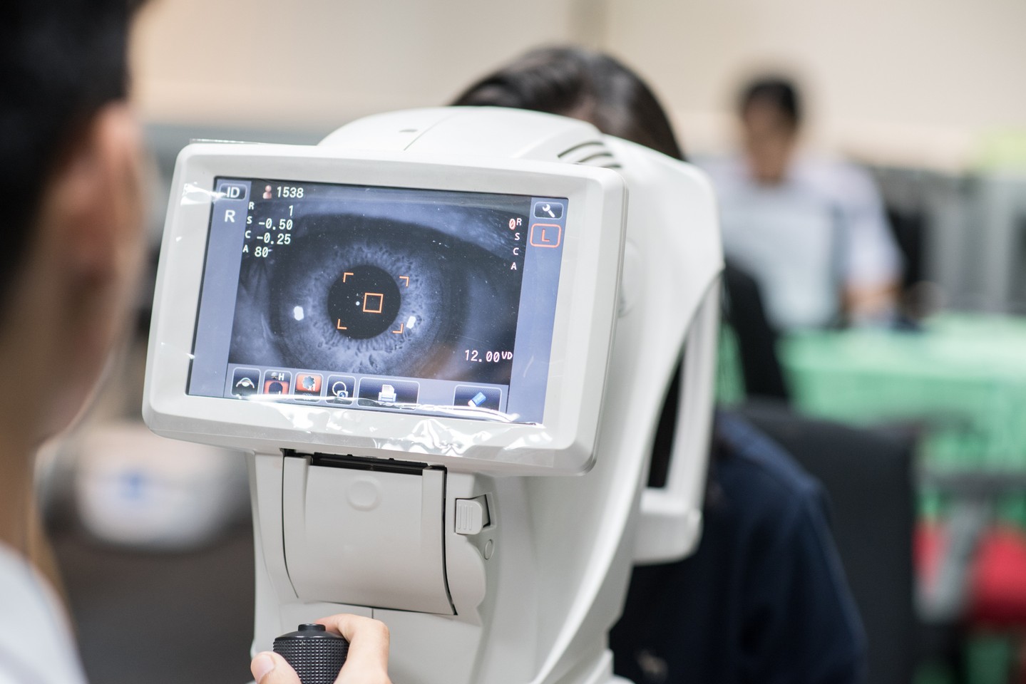
Retinal imaging refers to several advanced diagnostic technologies that create detailed pictures of the retina and surrounding structures inside the eye.
Unlike traditional photographs, these imaging systems provide physicians with highly detailed information about the layers of the retina, blood vessels, and other tissues that cannot always be evaluated with a standard eye examination alone. These tests are painless, non-invasive, and typically completed in just a few minutes.
Optical Coherence Tomography (OCT)
One of the most valuable tools in modern retina care is Optical Coherence Tomography, commonly known as OCT. OCT uses light waves to create high-resolution cross-sectional images of the retina. These images allow retina specialists to examine each layer of retinal tissue in remarkable detail. OCT is commonly used to diagnose and monitor:
- Age-related macular degeneration
- Diabetic macular edema
- Epiretinal membranes
- Macular holes
- Central serous retinopathy
- Other retinal disorders affecting the macula
Because OCT images can be repeated over time, physicians can compare scans from previous visits to determine whether a condition is stable, improving, or progressing.
Retinal Photography
Digital retinal photography captures detailed color images of the back of the eye. These photographs document the appearance of the retina, optic nerve, and retinal blood vessels, creating an important visual record that can be compared over future visits.
Retinal photography is especially helpful for monitoring diabetic eye disease, retinal hemorrhages, vascular disorders, and changes associated with aging.
Fluorescein Angiography
When additional information about retinal blood flow is needed, your physician may recommend fluorescein angiography.
During this test, a fluorescent dye is injected into a vein in the arm while specialized photographs are taken as the dye travels through the blood vessels of the retina.
This study allows retina specialists to identify leaking blood vessels, abnormal circulation, and areas of retinal damage that may not be visible during a standard examination. Not every patient requires this test, but it can provide valuable diagnostic information in selected cases.
Imaging Helps Guide Treatment
Retinal imaging doesn’t simply diagnose disease. It also helps physicians determine the most appropriate treatment and monitor how well that treatment is working.
By comparing imaging studies over time, retina specialists can evaluate whether medications are reducing retinal swelling, whether bleeding has resolved, or whether a retinal condition remains stable. This information allows treatment plans to be personalized for each patient’s individual needs.
A Better Understanding of Your Eye Health
One of the greatest benefits of modern retinal imaging is the ability to help patients better understand their diagnosis.
Seeing detailed images of the retina often makes it easier to understand how a condition is affecting vision and why certain treatments or follow-up visits are recommended. These conversations help patients become active participants in protecting their long-term eye health.
Technology Combined with Experience
While advanced imaging provides extraordinary detail, technology is only one part of exceptional retinal care.
Interpreting these images requires specialized training and clinical experience. Retina specialists combine advanced diagnostic technology with years of expertise to recognize subtle changes, diagnose complex diseases, and recommend the most appropriate treatment for each patient.
At Mississippi Retina Associates, advanced retinal imaging plays an important role in helping preserve vision through early detection, accurate diagnosis, and personalized care. If you have been referred to a retina specialist or are experiencing changes in your vision, comprehensive retinal imaging can provide valuable insight into your eye health and help guide your care.